Postmortem Imaging – Virtopsy
The Digital Revolution in Forensic Autopsy Imagine a forensic pathologist receiving a body after a suspicious death. Traditionally, the body would undergo an external and internal examination, including dissection, organ examination, sampling and documentation. Now imagine that, before making an incision, investigators can place the body inside a CT or MRI scanner and create a detailed three-dimensional digital record of the entire body—including fractures, foreign objects, gas collections, vascular structures and many soft-tissue findings. This is the basic idea behind Virtopsy, or virtual autopsy.
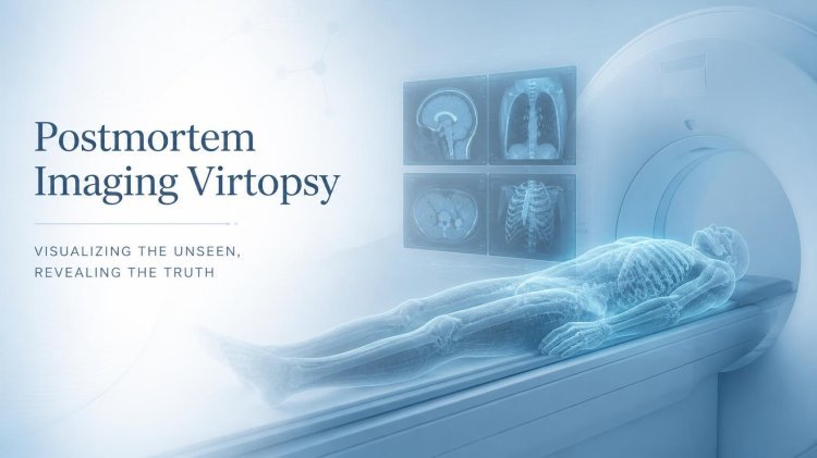
Virtopsy combines forensic medicine with postmortem CT, MRI, 3D surface scanning, image processing and sometimes angiography or image-guided sampling. The modern concept developed from the Virtopsy Project associated with the University of Bern in Switzerland in the late 1990s.
1. What is Virtopsy?
The word Virtopsy comes from:
Virtual + Autopsy = Virtopsy
It refers to the use of advanced imaging and computer-based techniques to examine a deceased person without relying solely on conventional dissection.
The central technologies include:
- Postmortem Computed Tomography (PMCT)
- Postmortem Magnetic Resonance Imaging (PMMRI)
- Postmortem CT Angiography (PMCTA)
- 3D surface scanning
- Photogrammetry
- Magnetic Resonance Spectroscopy (MRS)
- 3D image reconstruction
- Image-guided biopsy/sampling
Recent reviews describe PMCT as a major component of modern forensic imaging, while MRI provides complementary information, particularly for soft tissues and the central nervous system.
2. Why was Virtopsy developed?

Traditional autopsy remains extremely important, but it has some limitations.
A conventional autopsy:
- is invasive
- changes the condition of the body
- involves human interpretation
- can make later reassessment difficult
- may be unacceptable or problematic in some cultural or religious contexts
- cannot always preserve every finding in a readily reviewable 3D format
Virtopsy attempts to address some of these problems by creating a digital record of the body before destructive examination.
One major advantage is that the digital dataset can be stored and re-examined later by another forensic expert.
3. How does a Virtopsy investigation work?
A simplified workflow is:
Deceased body
⬇️
External documentation
⬇️
3D surface scanning
⬇️
Postmortem CT
⬇️
Postmortem MRI, if required
⬇️
Special imaging / angiography
⬇️
3D reconstruction
⬇️
Forensic interpretation
⬇️
Comparison with conventional autopsy, toxicology, histology and other evidence
⬇️
Final forensic opinion

4. Postmortem CT – The backbone of Virtopsy
Computed Tomography (CT) uses X-rays to create cross-sectional images of the body.
In postmortem CT, the deceased person is scanned after death.
The scanner produces a large number of thin slices that can be reconstructed into different views.
For example, the examiner can examine:
Axial view
A cross-section through the body.
Coronal view
A front-to-back oriented reconstruction.
Sagittal view
A side-oriented reconstruction.
3D reconstruction
A three-dimensional representation of anatomical structures.
Modern PMCT protocols can use high-resolution acquisitions and multiple reconstructions because radiation exposure to a deceased person is not a concern in the same way as it is for living patients.
5. What can CT detect?
CT is particularly valuable for bones and high-density materials.
It can help identify:
- skull fractures
- facial fractures
- rib fractures
- spinal injuries
- pelvic fractures
- foreign bodies
- bullets and fragments
- dental structures
- gas collections
- some internal injuries
- skeletal trauma
- fracture patterns
This makes PMCT especially useful in trauma, firearm and unidentified-body investigations. A recent scoping review identified PMCT as particularly effective for complex osseous injuries, bullet trajectories and some characteristic findings in drowning cases.
6. Virtopsy in firearm injuries

This is one of the most impressive applications.
Suppose a body has a suspected gunshot injury.
Traditional examination may require careful dissection to determine:
- bullet path
- entry wound
- exit wound
- internal fractures
- bullet fragments
- projectile location
PMCT can provide a three-dimensional view of the projectile and associated skeletal damage.
For example:
Entry region
⬇️
Bone damage
⬇️
Projectile trajectory
⬇️
Bullet/fragments
The 3D dataset can then be manipulated to examine the trajectory from different perspectives.
However, imaging is interpreted together with the external examination, scene findings, autopsy and other evidence—it does not independently establish every aspect of a shooting event.
7. Postmortem MRI
While CT is excellent for bones and many traumatic findings, MRI is particularly useful for soft tissues.
MRI can provide detailed information about:
- brain
- spinal cord
- muscles
- organs
- soft-tissue injuries
- hemorrhage
- some cardiac structures
- neurological abnormalities
Research comparing modalities has found PMMRI particularly valuable for soft-tissue and neurological assessment
8. CT vs MRI in Virtopsy
| Feature | Postmortem CT | Postmortem MRI |
|---|---|---|
| Bone | ⭐⭐⭐⭐⭐ | ⭐⭐ |
| Fractures | Excellent | Good |
| Bullet/metal detection | Excellent | Limited/affected by artefacts |
| Brain | Good | Excellent |
| Soft tissue | Good | Excellent |
| 3D bone reconstruction | Excellent | Less suitable |
| Vascular imaging | With special PMCTA | Possible with appropriate techniques |
| Speed | Relatively fast | Usually slower |
| Whole-body trauma | Excellent | Useful complement |

9. Postmortem CT Angiography – PMCTA
One limitation of ordinary CT is that the body's blood circulation has stopped.
Therefore, conventional clinical CT angiography cannot simply be transferred unchanged to a deceased body.
Postmortem CT angiography (PMCTA) uses specialized techniques to introduce a contrast agent into the vascular system.
This can help visualize:
- arteries
- veins
- vascular injuries
- vessel abnormalities
- some sources of bleeding
Research reviews identify PMCTA as particularly useful for investigating vascular lesions and certain natural-death cases.
10. 3D Surface Scanning
Virtopsy is not only about looking inside the body.
The external surface can also be digitally captured.
3D surface scanning can document:
- wounds
- bruises
- abrasions
- lacerations
- burns
- patterned injuries
- body morphology
- facial features
A digital 3D model can preserve the external appearance for later examination.
The original Virtopsy approach integrated CT/MRI with 3D surface documentation.
11. Photogrammetry
Another technique is photogrammetry.
Multiple photographs are taken from different angles.
Specialized software then uses these images to construct a three-dimensional representation.
For example:
???? Photograph 1
???? Photograph 2
???? Photograph 3
???? Photograph 4
↓
???? Photogrammetry processing
↓
???? 3D surface model
This can be particularly useful when documenting external injuries or the overall appearance of remains.
The two techniques are therefore complementary rather than competitors.

12. 3D Reconstruction
This is where Virtopsy becomes especially powerful.
Instead of looking at hundreds or thousands of individual CT slices, the examiner can create a 3D reconstruction.
For example, a skull can be digitally reconstructed and rotated.
The examiner may then examine:
- fracture lines
- bone displacement
- foreign bodies
- dental structures
- anatomical relationships
13. Virtopsy and identification of unknown bodies
Imagine investigators recover an unidentified body.
Traditional identification methods may include:
- fingerprints
- dental comparison
- DNA
- anthropological examination
Postmortem imaging can provide additional information.
CT can document:
- dental structures
- previous fractures
- surgical implants
- prostheses
- anatomical abnormalities
- healed injuries
These findings may then be compared with ante-mortem medical or dental records.
Forensic imaging has long been used in identification, including dental comparisons and reconciliation of ante-mortem and postmortem imaging.
14. Virtopsy in forensic odontology
Dental structures are particularly resistant to decomposition and can be highly valuable for identification.
CT can document:
- teeth
- roots
- jaw structures
- restorations
- implants
- dental morphology
A 3D reconstruction may help a forensic odontologist examine the dental structures without immediately performing extensive physical intervention.
This can be especially useful when dealing with severely damaged or decomposed remains.
15. Virtopsy in trauma investigation
Suppose a person dies after a fall.
The body may have:
- skull fractures
- rib fractures
- vertebral injuries
- pelvic fractures
- internal bleeding
- soft-tissue trauma
PMCT can rapidly provide a whole-body overview.
The examiner can then focus on areas requiring more detailed investigation.
This is one reason postmortem imaging can work as an early investigative map before or alongside conventional autopsy.
16. Virtopsy and drowning investigations
Drowning is a particularly challenging forensic diagnosis because there is often no single finding that independently proves drowning.
Postmortem imaging can provide useful information, including certain patterns involving:
- lungs
- airways
- fluid/gas distribution
- sinuses
- skeletal injuries
- associated trauma
Recent reviews identify PMCT as useful for detecting characteristic findings in some drowning investigations, but imaging findings must be interpreted within the entire case context.
This is an important principle:
Virtopsy supports forensic diagnosis; it does not turn a complex diagnosis into a simple one.

17. Virtopsy and decomposition
Postmortem imaging can also document the body when decomposition has begun.
CT can provide information about:
- skeletal structures
- gas distribution
- foreign objects
- remaining anatomical structures
However, decomposition itself creates imaging changes that can sometimes complicate interpretation.
Therefore, the examiner needs specific knowledge of postmortem changes.
18. Virtopsy and time since death
There has been research into using imaging and advanced techniques to estimate the postmortem interval (PMI).
Potential approaches include:
- decomposition-related imaging changes
- MRI
- magnetic resonance spectroscopy
- biochemical analysis
- temperature measurements
- computational models
Magnetic resonance spectroscopy has been investigated for biochemical changes occurring after death, but PMI estimation remains complex and should not be presented as a simple "CT tells the exact time of death" technique.
19. Magnetic Resonance Spectroscopy — MRS
MRS goes one step further than conventional MRI.
Instead of primarily creating anatomical images, it can provide information about chemical metabolites in tissues.
In forensic research, MRS has been investigated for studying postmortem biochemical changes and their potential relationship with postmortem interval.
This remains more specialized and research-oriented than routine PMCT.
20. Micro-CT
For very small structures, micro-computed tomography (micro-CT) can provide extremely high-resolution imaging.
It can be useful for:
- small bones
- teeth
- bullets
- tissue samples
- anatomical structures
- archaeological/anthropological specimens
Earlier Virtopsy research described micro-CT systems capable of imaging small samples at very high resolution.
21. Virtopsy in child and fetal death investigations
Postmortem MRI has become particularly important in some fetal, neonatal and pediatric investigations.
Why?
Because very detailed soft-tissue information can sometimes be obtained without extensive dissection.
This can be valuable when parents have concerns about invasive examination.
Current reviews identify pediatric and fetal investigations as areas where postmortem MRI has particular relevance

Virtopsy and conventional autopsy
| Conventional Autopsy | Virtopsy |
|---|---|
| Physical examination | Digital examination |
| Invasive | Non-invasive/minimally invasive imaging |
| Direct tissue examination | Imaging-based examination |
| Histology possible directly | Requires sampling for histology |
| Toxicology sampling possible | Can guide targeted sampling |
| Strong for many pathological findings | Strong for 3D anatomy and trauma documentation |
| Can alter the body | Preserves digital record before intervention |
| Human interpretation | Digital, reproducible datasets |
| Re-examination may be limited | Dataset can be reviewed repeatedly |
But Virtopsy should not automatically be considered a complete replacement for conventional autopsy.
Some diseases, toxicological conditions and microscopic findings cannot be adequately established from imaging alone.
Virtopsy and courtroom presentation
3D reconstructions can also help explain complicated injuries.
Instead of showing a court dozens of CT slices, an expert may use:
- axial images
- sagittal images
- coronal images
- 3D bone reconstructions
- vascular reconstructions
- annotated models
For example, a complex skull fracture can be much easier to understand through a rotating 3D model.
However, the original imaging data and reconstruction methodology must remain available, and the presentation should clearly distinguish observed findings from interpretations.
Role of Artificial Intelligence
The next stage of Virtopsy is likely to involve AI-assisted forensic imaging.
AI could potentially help with:
- fracture detection
- segmentation of organs
- identification of foreign objects
- bone analysis
- 3D reconstruction
- vascular segmentation
- image classification
- comparison of ante-mortem and postmortem images
- automated measurements
A future system might look like:
PMCT
↓
AI identifies possible fracture
↓
AI segments fracture
↓
3D reconstruction
↓
Forensic pathologist verifies finding
The human expert remains essential because forensic conclusions require context, validation and interpretation.
Recent literature describes increasing integration of computational science and AI with postmortem imaging.
Follow cyberdeepakyadav.com on
Facebook, Twitter, LinkedIn, Instagram, and YouTube
What's Your Reaction?