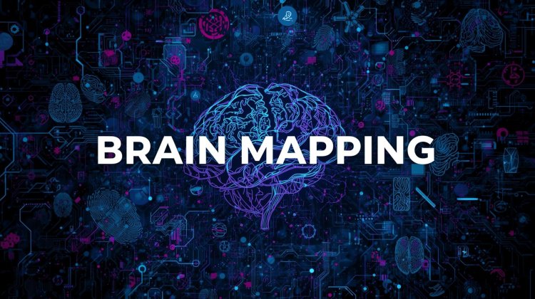
Brain Mapping: A Window into Criminal Memory
This technique is used to determine scientifically what information is in or what information is not stored in the brain. It measures the response to the visual and audio stimulus. Stimulus is a thing or event that evokes a specific functional reaction in organ or tissue.
It measures the electrical brainwave response towards phrases or pictures that are presented on a computer screen.
The study and visualisation of brain activity through the use of sophisticated neuroimaging and electrophysiological instruments is known as brain mapping. Knowing which areas of the brain light up in response to particular stimuli is helpful. It is used to determine if a suspect has experiential knowledge—the real recollection of an incident or crime—in forensic circumstances.
INVENTION
Foundation of the Idea
1965 – Samuel Sutton (USA):
-
-
Discovered the P300 brainwave (a positive peak in brain activity about 300 milliseconds after recognizing something familiar).
-
This discovery became the scientific basis for brain fingerprinting.
-
1990s – Dr. Lawrence A. Farwell (USA):
-
An American neuroscientist and inventor.
-
Developed Brain Fingerprinting by combining EEG technology with the P300 brainwave principle.
-
Introduced a stronger response called MERMER (Memory and Encoding Related Multifaceted Electroencephalographic Response), which is more accurate than P300 alone.

Components of MERMER
-
P300 Wave
-
A positive electrical peak that occurs about 300 milliseconds after the brain recognizes something familiar.
-
-
Additional Brain Responses
-
MERMER also includes a late negative response and a frequency change in the brain’s electrical activity, making it more detailed than P300 alone. When brain recognise something then there is increase in nervous activity.
-
Techniques Used in Brain Mapping
-
Electroencephalography (EEG):
-
Records electrical signals from brain neurons.
-
Useful for identifying abnormal patterns of recognition.
-
-
Functional Magnetic Resonance Imaging (fMRI):
-
Measures blood oxygen level changes in the brain.
-
Shows which areas become active when recognizing information.
-
-
Positron Emission Tomography (PET):
-
Measures glucose metabolism in brain cells.
-
Active regions consume more energy.
-
-
Brain Electrical Oscillation Signature Profiling (BEOS):
-
Special forensic tool developed in India.
-
Distinguishes between:
-
Experiential knowledge (suspect was at the crime scene)
-
Semantic knowledge (suspect just heard about it).
-
-

Procedure
-
The subject is made to sit in a relaxed position with electrodes attached to their scalp.
-
Crime-related stimuli (words, pictures, sounds) are shown.
-
Brain responses are recorded and analyzed by computer.
-
Report is generated indicating if the person had prior experiential knowledge.
Types of Brain Waves
-
Delta Waves (0.5 – 4 Hz)
-
Slowest brain waves.
-
Seen during deep sleep and unconscious states.
-
Important for healing and regeneration.
-
-
Theta Waves (4 – 8 Hz)
-
Associated with drowsiness, light sleep, deep relaxation, daydreaming.
-
Seen in meditation and creative states.
-
Also appears in children more than adults.
-
-
Alpha Waves (8 – 13 Hz)
-
Appear when a person is relaxed but awake.
-
Typical when eyes are closed and the mind is calm.
-
Related to creativity, relaxation, and reduced stress.
-
-
Beta Waves (13 – 30 Hz)
-
Associated with active thinking, problem-solving, concentration, alertness.
-
Too much beta activity = anxiety, stress.
-
-
Gamma Waves (30 – 100 Hz)
-
Fastest brain waves.
-
Linked to memory, learning, information processing, and perception.
-
Strong gamma activity = high-level thinking and consciousness.
-
Brain Waves and Color Coding (Red, Green, Blue)
In Brain Mapping (especially EEG/BEOS or neurofeedback systems), brain wave activity is displayed on a computer screen using colors. These colors indicate the level of brain activation:
Red (High Activity / Abnormal)
-
Shows over-activation of a brain region.
-
Means the neurons in that area are firing excessively.
-
Could indicate stress, anxiety, hyper-alertness, or abnormal recognition (in forensic cases: suspect may be recognizing crime-related information strongly).
Green (Normal / Balanced Activity)
-
Represents normal or healthy brain activity.
-
Indicates that the brain region is functioning within the expected frequency range.
-
In forensic mapping → green means no unusual recognition/memory trace.
Blue (Low Activity / Underactive)
-
Shows low or reduced activation of brain cells.
-
Could mean drowsiness, reduced attention, or suppression of memory recall.
-
In forensic use → blue may suggest the person does not recognize or is not processing the crime-related stimulus.
Red and blue waves are closely related or stimulated close to each other it means that the person known the information of crime.

Stages of brain mapping
-
Preparation – subject setup with EEG.
-
Baseline Recording – normal brain activity noted.
-
Stimulus Presentation – crime-related words/images shown.
-
Brain Wave Recording – EEG captures responses.
-
Analysis – software checks for BEOS signatures.
-
Report – conclusion: experiential knowledge present/absent.
Applications
-
Terrorism investigations (e.g., identifying trained terrorists).
-
Murder and sexual assault cases.
-
Espionage and intelligence investigations.
-
Verification of witness statements.
First Use in Courts
-
India became the first country to use brain mapping (BEOS) in criminal investigations.
-
Example: The Aarushi Talwar double murder case (2008), where BEOS tests were used on suspects.
-
Though controversial, it showed that brain mapping had entered forensic practice.
Follow cyberdeepakyadav.com on
Facebook, Twitter, LinkedIn, Instagram, and YouTube
What's Your Reaction?